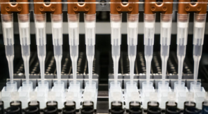
NGS Panels
Next-generation sequencing (NGS) technology has completely changed the field of genomic medicine by allowing researchers to better comprehend human diseases and pinpoint their genetic underpinnings. These initiatives have sparked the creation of effective preventive strategies and assisted medical professionals in developing pharmaceuticals that are particularly formulated to treat a variety of illnesses without side effects.
We offer a broad selection of NGS panels that have been created with an emphasis on offering diagnosis and prognosis choices for the treatment and prevention of a number of hereditary and somatic malignancies, cardiovascular, pulmonary, and neurological disorders
What is Whole genome sequencing (WGS)?
Every living creature has a unique genetic code, or genome, made up of nucleotide bases, including bacteria, plants, and animals (A, T, C, and G). If you know the order of the bases, you can identify an organism’s unique DNA pattern. The procedure of determining the bases’ order is known as sequencing. A laboratory technique known as whole genome sequencing can be used to establish the base order of an organism’s genome in a single step.
What is the process of whole genome sequencing?
The four main steps that scientists use to accomplish whole genome sequencing are as follows:
- DNA shearing: The DNA, which is composed of millions of bases (As, Cs, Ts, and Gs), is first divided into manageable pieces that can be read by a sequencing machine using molecular scissors.
- DNA bar coding: The addition of small DNA tags or bar codes allows researchers to identify whose bacterium a sheared DNA fragment belongs to. The way a grocery store’s bar code identifies a product is similar to how this does.
- DNA sequencing: The bar-coded DNA of several bacteria is combined and put into a DNA sequencer. The sequencer identifies the A, C, T, and G bases, or structural components, of each bacterial sequence. The sequencer uses the bar code to keep track of which bases belong to which bacteria.
- Data analysis: Researchers use computer analysis methods to analyze sequences from distinct microorganisms and identify differences. The degree of differences among the bacteria can be used to estimate their degree of similarity and the likelihood that they are linked to the same outbreak.
Exome sequencing, commonly referred to as whole exome sequencing (WES), is a genetic technique for sequencing each gene’s protein-coding region (known as the exome). There are two parts in this process: the first is to only select DNA sequences that code for proteins. These regions are called exons, and there are about 180,000 of them in the human genome, or about 30 million base pairs, or about 1% of the entire genome. The next step is to sequence the exonic DNA using a high-throughput DNA sequencing method.
Why Whole Exome Sequencing?
This approach seeks to identify genetic variants that alter protein sequences as opposed to whole-genome sequencing. Because these mutations can be the source of Mendelian and common polygenic illnesses, such Alzheimer’s disease, whole exome sequencing has been employed in both academic research and clinical diagnosis.
- Allows entire exon coverage to focus on medically relevant genomic regions, such as untranslated and known disease-associated regions (UTRs).
- Increases the likelihood that variants may be discovered using next-generation sequencing (NGS) technology, particularly rare and low-frequency mutations.Provides a less expensive alternative to WGS and eliminates the necessity for complete genome sequencing.
Transcriptome
The term “transcriptome” refers to the whole collection of messenger RNA, or mRNA, molecules that a given organism expresses. The term “transcriptome,” or transcriptome, refers to the range of mRNA transcripts produced in a particular cell or tissue type. Contrary to the genome, which is characterized by its stability, the transcriptome actively changes. In reality, an organism’s transcriptome varies depending on a wide range of factors, such as its environment and stage of development.
The microarray is a laboratory technique that can be used to investigate the transcriptome. Microarrays can be used to simultaneously assess the expression of thousands of genes in addition to generating gene expression profiles, which describe changes in the transcriptome in response to a specific condition or medication.
Whole transcriptome sequencing (WTS) enables V-Labs to gain greater understanding of the RNA patterns of patient malignancies. WTS provides extraordinarily thorough coverage (coverage) and outstanding resolution (depth) into the dynamic dynamics of the transcriptome.
With WTS, it is feasible to analyze gene expression, find gene fusions, and find RNA splice variants with just one simple test. The test can identify fusions regardless of DNA breakpoints and can detect unusual or novel fusion events better than targeted RNA sequencing or DNA-based techniques. Because whole transcriptome sequencing provides comprehensive exon coverage and gathers virtually all potential fusion partners, it can reliably detect different forms of expressed gene fusions while reducing false positives brought on by non-expressed rearrangements.
With Whole Exome Sequencing (WES), Whole Transcriptome Sequencing (WTS), extensive proteome testing services, and cutting-edge machine-learning capabilities, V-Labs molecular profiling provides the most comprehensive and therapeutically applicable molecular profile for cancer patients on the market.
Advantages of RNA Analysis via Whole Transcriptome Sequencing
Chromosomal rearrangements.
- DNA sequencing in formalin-fixed materials can detect some chromosomal rearrangements, but it can be difficult to find breakpoints within large regions of repeating intronic sequence when using short sequence reads.
- If a fusion gene is expressed in mRNA, DNA sequencing CANNOT show it.
- DNA sequencing is unable to find read-through fusions.
Variants Transcripts
- Because of variation mRNA splicing, variant mRNA transcripts cannot be directly detected by DNA sequencing.
- MI Transcriptome RNA sequencing WILL find variant transcripts by simply looking at the spliced mRNA.
- When examining biological processes, RNA sequencing yields more useful data; MI Transcriptome will offer an estimation of the levels of gene expression for use by pathway analysis software.
- Targeted Panel
Using targeted gene sequencing panels, one can examine certain mutations in a sample. Focused panels include a particular group of genes or gene regions that are either known to be or are most likely to be associated with the condition or characteristic being studied. Pre-selected gene panels are available for purchase, or custom panels can be created to incorporate specific genomic areas.
Due to its scalability, speed, and resolution, next-generation sequencing (NGS) enables the evaluation of particular genes of interest. When compared to performing each experiment independently, many samples can be used to study many genes at once, saving time and money. Targeted gene sequencing produces a smaller, more manageable data set than more exhaustive techniques like whole-genome sequencing while also facilitating analysis.
We offer single, double, or triple variant sequencing for any of the genes on our test menu.
– Prenatal testing
– Non invasive Prenatal testing (NIPT)
– Targeted testing (PCR and/or FISH) of relatives of patients who had copy number testing (del/dup studies via aCGH or chromosomal microarray studies (CMA)) at Prevention Genetics is available.
Benefits of Targeted Gene Sequencing
- High-depth sequencing of crucial genes or regions of interest enables the discovery of rare variations (500–1000 or higher).
- Offers cost-effective results for research on genes associated with disease.
- Provides precise, comprehensible data and has the ability to detect changes with low allele frequencies (down to 5%).
- Makes it possible for unique or inherited causative mutations to be precisely detected in a single experiment.
- Pharmacogenomics / Genetics
Why do we use Pharmacogenomics (“PGX”)?
V-Labs America’s mission is to change and improve people’s lives by giving them and their doctors the information and therapeutic outcomes they need through pharmacogenomic testing.
Why shouldn’t a person’s medications take into account the variations in their genetic make-up and physical structure? Genetic information can be used to forecast how a person’s body will respond to and metabolize drugs. How a drug is metabolized can have a big impact on whether it is therapeutically effective or harmful. For instance, a medicine may not work if the body digests it too rapidly or too slowly. If a physician doesn’t sure which medication will be the best choice, they may have to choose a prescription based on prior experience or drug prescribing data in the hopes that the patient’s body will respond well to it. Pharmacogenomic research utilized in personalized medicine helps to reduce some of the mystery around drug effectiveness. Utilizing cutting-edge technologies and bioinformatics tools, V-Labs America screens and assesses the role of DNA mutations that are likely to affect drug pharmacokinetics (drug absorption, distribution, metabolism, and elimination) and pharmacodynamics (therapeutic target) as well as their impact on bioavailability, therapeutic response, and safety. These genetic variations will be utilized to assess the likelihood of negative drug reactions as well as the likelihood that a specific treatment would be beneficial on a specific person.
What is Pharmacogenomics (“PGX”) ?
Pharmacogenomics is the study of how a person’s DNA affects how they react to medications.
- Pharmacogenomic testing has the potential to improve clinical outcomes and increase the financial value of the healthcare system. Making pharmacogenomics available to physicians and patients can result in significant cost savings, according to a number of compelling studies.
- V-Labs America specializes in pharmacogenomics lab testing, which identifies genetic variants that affect the metabolism of therapeutic medications and the possibility of unfavorable results.
Having a list of the patient’s current medications as well as a list of potential drug-drug, gene-drug, and drug-gene-drug interactions may help a doctor better understand why a patient would respond to a medication in a particular way.
To help uncover mutations that potentially affect a person’s metabolism and/or response to medication, V-Labs America, a pharmacogenomics lab, tests more than 30 genes. A pharmacist at V-Labs America reviews a Personalized Medication Report following the pharmacogenomic analysis, which was conducted using a simple cheek swab sample, in order to look for potential gene-medication interactions, medication-medication interactions, and medication-gene-medication interactions. A tailored medication guide is also available, which includes medications by specialty under the headings “Use as Directed,” “Use with Care,” and “Suggest Alternate Medicine.” This list may be used by a prescriber to help them choose new or different medications for their patient. The report is a useful resource for helping patients and medical professionals decide on the best course of action.
Test Menu for Medication Management
Cytochrome P450 Enzymes: The Cytochrome P450 enzyme system, one of many metabolic systems, controls a substantial portion of the metabolism of medicines. A substrate is a material that an enzyme breaks down. Once a medication has been digested, a certain enzyme becomes “active” in the body, meaning some pharmaceuticals are also prescriptions for that enzyme. Drugs can be metabolized by people differently because of variations in the relevant enzymes. There are four classifications for each enzyme’s metabolic rate: normal metabolism, quick metabolism, intermediate metabolism, and poor metabolism.
- Toxicology
The study of chemicals, physical agents, or biological agents that influence living beings and the ecosystem, as well as methods to avoid or mitigate these adverse effects, is known as toxicology. Toxicology is an applied science with numerous subfields of study. Toxicology requires integrating knowledge from many different fields of expertise. Most toxicologists carry out the following types of work in order to assess and comprehend how toxins affect living systems:
- Developing mechanistic understanding of effects
- Ensuring safer chemical products
- Developing safer drugs and medicines
- Determining risks from chemical exposures
- Developing treatments for chemical exposures
- Ensuring a safe food and water supply
Why Toxicology Test is needed?
A drug test may be necessary for a number of reasons. It can be essential for your new job, academic program, or insurance coverage. One is required by many sporting organizations of its participants.
- Are in treatment for drug addiction
- Show signs of substance abuse
- Have problems with your mental health
- Have been taking a controlled drug for a long period of time
The law may also necessitate a drug test. You might need to go on one if you’ve been accused of a crime. The same is accurate for many parolees.
Chemistry
To evaluate a person’s general health, chemistry panels are groupings of tests that are routinely ordered. They assist in determining things like the body’s electrolyte balance and/or the health of several crucial organs. The testing requires a blood sample, which is normally drawn from a vein.
Some panel titles, as well as the number and type of tests they contain, have been standardized nationally. Common chemistry panel we offer includes:
- Basic Metabolic Panel (BMP) – usually contains 8 tests, which is typically included in the CMP. It gives details on your blood glucose level, electrolyte balance, acid/base status, and present state of your kidney and respiratory systems.
- Comprehensive Metabolic Panel (CMP): This panel typically consists of 14 tests. With the inclusion of your liver’s condition and significant blood proteins, it offers the same details as the BMP.
- Electrolyte Panel: useful for detecting problems with the body’s electrolyte and fluid balance
- Lipid Panel: used to determine a person’s risk of developing cardiovascular disease
- Liver Panel: used to screen for, find, evaluate, and keep track of acute and chronic liver inflammation (hepatitis), liver illness, and/or damage.
- Renal Panel: contains tests such as creatinine, albumin, BUN, eGFR to evaluate kidney function
- Thyroid Function Panel: used to assess thyroid gland activity and identify thyroid diseases
Hematology
The significance of quick and complete responses is underscored by the genetic component that characterizes the majority of hematologic diseases. Genetic variants associated with hematological illnesses can be found in more than 300 different genes. Our genetic tests draw on our bio/databank that is especially devoted to rare diseases as well as our own internal medical expertise to provide precise and accurate diagnoses, prognoses, and potential treatment options, where necessary.
The V-Labs America Laboratory performs a wide range of basic and advanced hematology tests on whole blood, serum, urine, cerebrospinal fluids, and other body fluids.
Specific areas include:
- Standard hematology examinations, such as bone marrow staining, complete blood count, blood differential, and smear morphology.
- Regular coagulation tests (PT and PTT).
- Advanced hematology examinations, such as flow cytometry exams for the detection of lymphomas, leukemias, plasma cell neoplasms, systemic mastocytosis, paroxysmal nocturnal hemoglobinuria, and other lymphoproliferative diseases.
- The electrophoresis of hemoglobin.
- More complex tests for coagulation, include ristocetin titration, antiphospholipid antibody testing, platelet antibody and aggregation, hypercoagulability studies, coagulation factor and inhibitor investigations, and others.
- Cell counts, bodily fluid analysis, and urine analysis
- Immunoassay
A biochemical test called an immunoassay (IA) employs an antigen or an antibody to determine if or how much of a macromolecule or a small molecule is present in a solution (sometimes). The process is based on the idea that particular antigens attach to particular recently introduced antibodies, inducing an immunological response. This alludes to specificity since antibodies have a high degree of specificity for a wide range of analytes, including hormones, nucleoproteins, and peptides. Antigen-antibody complexes can successfully identify analytes when utilized in immunoassay assays if the reagents are pure. When the questioned analyte is present, a color shift happens. The amount of the targeted chemical in the test solution is indicated by the visible color.
ELISA Immunoassay Testing:
In an enzyme-linked immunosorbent assay, an ELISA reader is used to identify the color contrast on the ELISA plate. Spectrophotometry, fluorescence, and luminescence are used by the microplate reader. It calculates the amount of light at a specific wavelength that an analyte absorbs, reflects, or emits. The standards of interpretation are based on the target analyte and the negative control being present at preset quantities in ELISA plates and tube kits. The predetermined standards for the semi-quantitative readout are displayed as a range of blue colors that the ELISA reader concentrates on. There is also the option of a quantitative interpretation. To simultaneously measure the optical densities of all the samples and standards, it is necessary to add microwells to a plate reader.
Chemiluminescent Immunoassay Testing:
Using chemiluminescent substances and an antigen sample, molecular material is identified during this immunoassay test. It functions according to the core principle of antigen-antibody specificity. The way the enzymes function is by changing a substrate into a unique product, which causes the emission of a certain color of light. The luminescence indicates the presence of an antigen.
In ELISA and membrane assays, lower limits of detection can be attained by employing chemiluminescent substrates, which are more sensitive than other substrates. Due to the increase in sensitivity, proper selection of the substrate, antibody/antigen titers, and blockers is required during the optimization phase.
Lateral Flow Immunoassay Tests:
Lateral flow devices are immunoassay diagnostic equipment used to identify the presence of target analytes in test samples by introducing particular antibodies and allowing fluid to flow down the strip from one end to the other. The immunoassay test strip’s nitrocellulose membrane houses a sample pad and an absorbent pad, often known as a wicking pad. The applied fluid is absorbed by the wicking pad through capillary action. Separate lines of an antigen-specific antibody and an anti-conjugated antibody are seen on the nitrocellulose membrane. As it moves from the sample pad to the absorbent pad, the fluid interacts with the two lines—a control and a test line. The lines are the areas of detection. When the applied sample fluid contains the desired antigen, the analyte first attaches to the antigen-specific antibody, resulting in the formation of a visible line that denotes a positive result. When the fluid encounters the anti-conjugated antibody in the control line after passing through the membrane, a second line is created. The test is functioning properly, as evidenced by the second line. In the event that the second line doesn’t expand, researchers consider the experiment is invalid.
Advantages of immunoassays
Immunoassays provide a number of advantages over more established methods for analyzing food contamination, such as HPLC and GC. Immunoassays can provide a quick, simple, and inexpensive means of detection with sensitivity and specificity that are comparable to or better (in some circumstances) than the conventional methods. The quickness of the analysis is one of the main benefits of immunoassay.